
Fields of Interest
Light-Tissue Interactions
Light-Tissue Simulations and Research
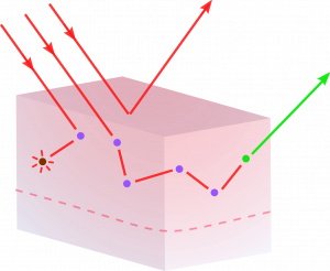
Through light interactions with tissues, we can discover the biological world around us. Most tissues and molecules can be interrogated using different light techniques, like one-, two- and three-photons excitation, FRET, FLIM, and super-resolution microscopy, among other exciting techniques that can make us discover essential properties of tissues, and see new, interesting phenomena. A lot of biological functions and molecules can be probed with optical techniques.
Medical Imaging Processing and AI
Multispectral Imaging
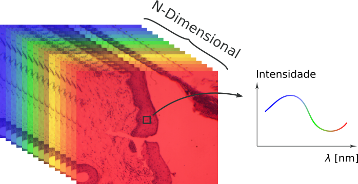
Multispectral imaging can capture the signature of a molecule’s absorption by the imaging of real-world scenarios. This technique can be used to probe tissues and molecules, from histology to widefield. A Hyperspectral system has been developed by us to acquire images in standard benchtop microscopes, and another setup was used to perform hemoglobin mapping in mice.
Artificial Intelligence in Image Classification
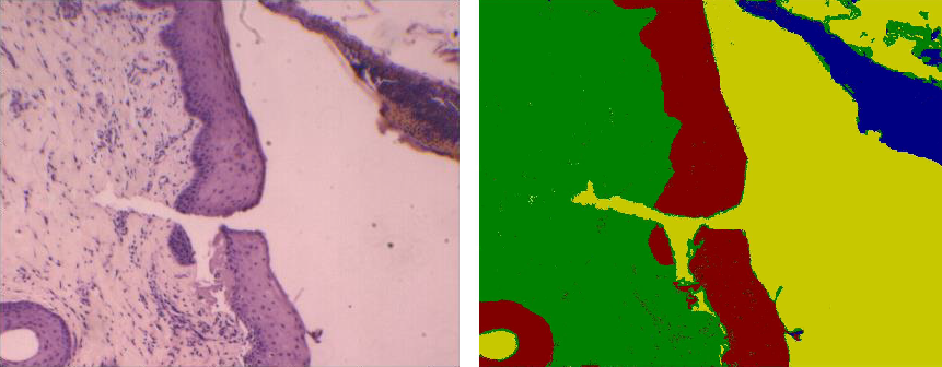
Artificial intelligence algorithms, like neural networks, are capable of analyzing images in histology slides and lesions to detect neoplasia regions of interest. In H&E stained histology, for example, the algorithm can be used to identify damaged regions, as in liver and skin lesions. This technique can also be used to classify and segment cell regions, evidencing cell conditions, or identify which cell types are in the image.
Optical and Electronic Instrumentation
Photodynamic Therapy Monitoring
Photodynamic Therapy (PDT) is a procedure to treat lesions and infections via deactivation with light, a photosensitizer, and oxygen. The photosensitizer is selective, can accumulate inside the tumor, and induce cell death by generating reactive oxygen species (ROS). The PDT monitoring in situ, in the clinical environment, is very important to afford both cure rate prediction and optimized protocol parameters delivering. The system developed in our group can make NIR fluorescence imaging and treatment of skin lesions at the same time, a step ahead in personalized medicine, which is pointed out in the literature as very important in photodynamic therapy development.
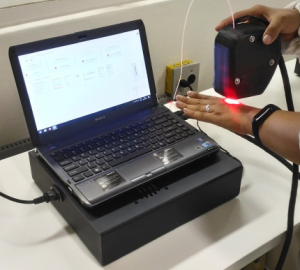
Digital Dermatoscope
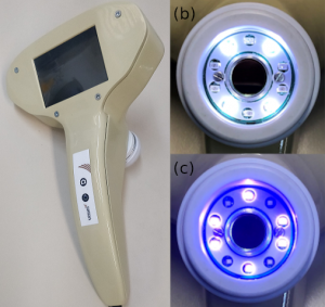
The digital dermatoscope is an instrument for skin and mucosal tissue visualization and quantification. It can help in monitoring treatments, like photodynamic therapy, or screening oral/mucosal lesions, which can present considerably more fluorescence than healthy mucosa. Here, we developed a dual-channel digital dermatoscope to acquire fluorescence and white light images over violet and white excitations.
Blood Superficial Perfusion
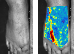
In a research developed in partnership with Rice University, a simple instrumentation is used to measure blood perfusion in lower members of diabetic patients, through imaging techniques. . It is intended to apply this instrumentation in a clinical test for the treatment of ulcers in diabetic feet. The project is being developed in collaboration with researchers from the Dept. of Electrical and Computing Engineering Department (ECE) at Rice University (Prof. Dr. Ashok Veeraraghavan and Prof. Dr. Ashutosh Sabharwal), with the Ph.D. students Amruta Pai and Akash Kumar Maity.
Fluorescence Microendoscope
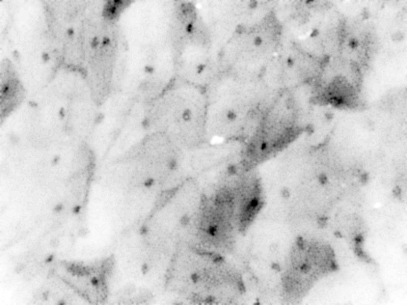
We also develop important techniques that can probe inside. In mucosa, techniques like fluorescence microendoscopy, can be used to probe interesting tissues, like the cervix and the mouth. We developed a dual-channel fluorescence microendoscope capable of capturing cells in real time. This can be used to help the diagnosis and procedure-guidances.